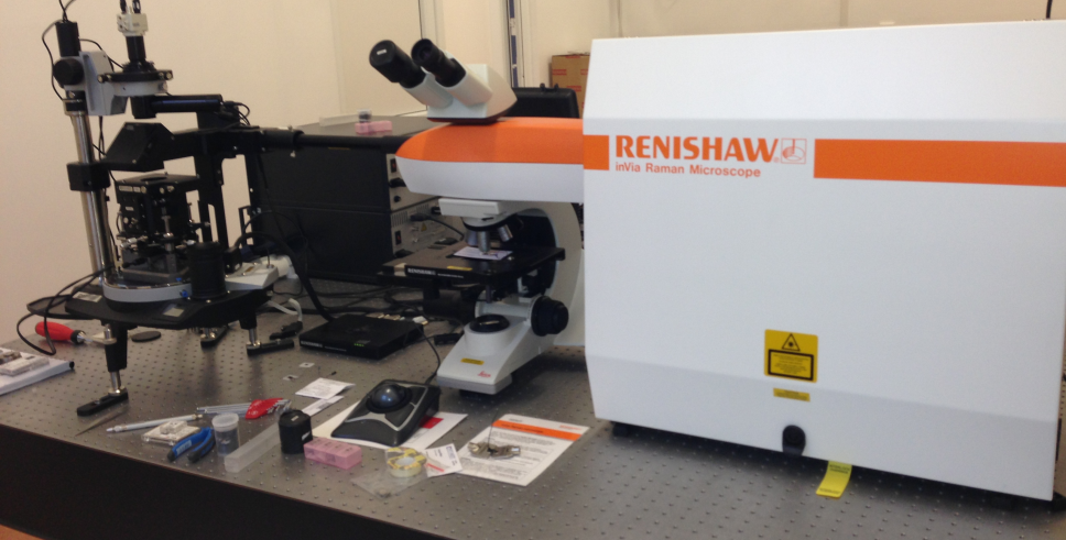
The Renishaw InVia Reflex Raman microscope includes a research-grade optical microscope (Leica) coupled to a high-performance Raman spectrometer. It is a new generation microscope that offers a powerful non-destructive and non-contact method of sample analysis. The instrument delivers both highly specific discrete analysis and information-rich images for a wide range of material types. It is perfectly suited for rapid, non-destructive analysis in life science, materials science, physics, and chemistry.
Reflection Raman microscopy enables physico-chemical analysis of elements and compounds with a spatial resolution of less than 1 mm (lateral) and less than 2 mm (depth). Raman shifts within 100 cm-¹ of the excitation frequency can be measured at excitation wavelengths of 325,442, 532, 633, 785 and 830 nm nm with a spectral resolution of 0.5 cm-1 in visible and 1 cm-1 in NUV and IR. Moreover, Raman shifts within 10 cm-1 of the excitation frequency can be measured at excitation wavelengths of either 532 nm or 633 nm with a spectral resolution of 0.5 cm-1. Spatial maps of Raman intensity over areas as large as several square cm can be made using an automated scan stage. In addition, our equipment offers the possibility of StreamLine Raman imaging which is a novel mapping technique that has reduced total mapping times to a level that is becoming clinically practicable. Thus, histological application of Raman mapping is now possible.
The equipment permits specialized applications like: Raman imaging, polarized Raman, resonant Raman, low-frequency Raman, photoluminescence, simultaneous Raman/AFM, Adhesion Force Microscopy, AFM Lithography and Nanomanipulation, Atomic Force Spectroscopy, Conductive Probe AFM, Scanning tunneling microscopy.
Renishaw Raman spectrometer: Six laser excitation sources covering the spectral range from UV to NIR: 325, 442, 532, 633, 785 and 830 nm
NTEGRA Spectra AFM microscope: The AFM microscope is directly coupled to the Raman spectrometer for co localized AFM and Raman imaging simultaneously on the same pixel) on non-transparent samples (upright configuration). The system is ready prepared for TERS experiments